|
|
Emphysematous Cystitis
General Considerations
- Acute inflammation of bladder mucosa and underlying muscle
- Diabetes mellitus, frequently poorly controlled, is present in 50-80% of cases
- Other predisposing conditions
- Chronic urinary tract infections
- Neurogenic bladder
- Bladder outlet obstruction
- Women are affected twice as often as men
- Gas-forming organisms most often found include E. coli and Enterobacter aerogens, with Clostridia and Candida species found on occasion
Clinical Findings
- Dysuria
- Frequency
- Hematuria
- Pneumaturia is rare
Imaging Findings
- Conventional radiography may show gas in the lumen of the bladder or in the wall
- It is usually curvilinear or mottled and may assume an oval shape with a horizontal long axis as opposed to air in the rectum which may also be oval shaped but has a vertical long axis
- If the gas is intraluminal, there may be an air-fluid level using a horizontal x-ray beam or on CT
- When adjacent to the nondependent mucosal surface, the air may have a cobblestone or “beaded necklace” appearance
- US may show diffuse bladder wall thickening and increased echogenicity
- There may be focal regions of high-amplitude echoes with posterior dirty acoustic shadowing
- CT is highly sensitive and may show other causes of bladder air such as a fistula from bowel secondary to a carcinoma or inflammatory disease such as diverticulitis or Crohn’s Disease
Differential Diagnosis
- Bladder instrumentation
- Vesicocolic or vesicovaginal fistulas
- Trauma
Treatment
- Treatment involves intravenous antibiotics and relief of any outlet obstruction.

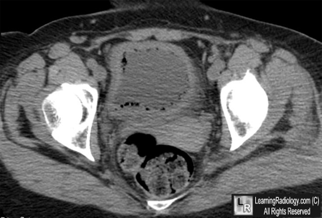
Emphysematous Cystitis. Axial, non-contrast enhanced CT scan of the pelvis demonstrates multiple small bubbles of air in the wall of the urinary bladder (white arrows). The bladder wall is thickened.
For this same photo without the arrows, click here
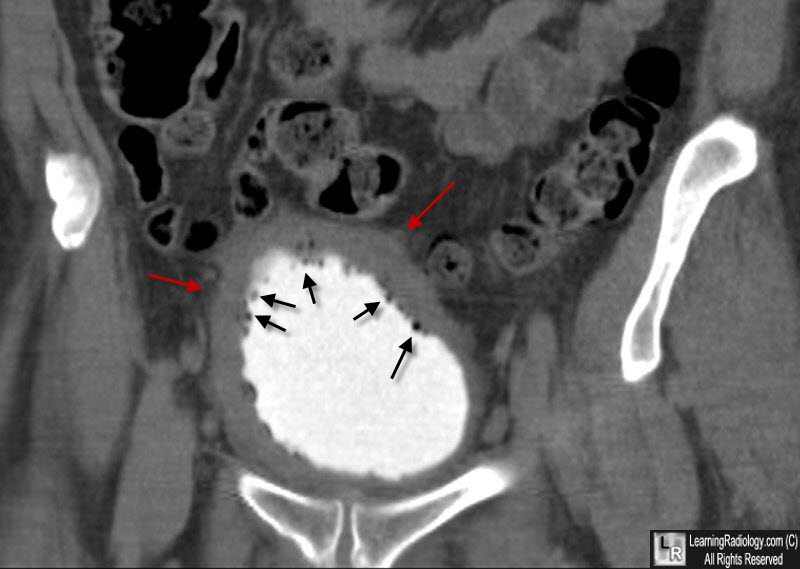
Emphysematous Cystitis. Coronal CT scan with contrast in the urinary bladder shows small bubbles of air (black arrows) in a markedly thickened bladder wall (red arrows).
Emphysematous Infections of the Abdomen and Pelvis: A Pictorial Review. DE Grayson; RM Abbott; AD Levy and PM Sherman. May 2002 RadioGraphics, 22, 543-561.
|
|
|
